| Genus: |
Brachysomus Schoenherr, 1823: 1124 |
| |
|
Brachysomus Schoenherr, 1823: 1124 (Type: Curculio echinatus Bonsdorff, 1785: 33)
Platytarsus Schoenherr, 1840 (Syn.)
The native range of the genus Brachysomus is limited to the northern Palaearctic Region. The only known exception is transpalaearctic B. echinatus (Bonsdorf), which was introduced in North America and Japan. The northern distribution boundary of B. echinatus in the Palearctic is passing across Karelia and Komi, while its southern boundary passes through Transcaucasia (Fig. 476); these boundaries mark the northern and, along with Brachysomus tannourinensis Szénasi & Németh from Lebanon, mark the southern limits of the range of the entire genus Brachysomus. A somewhat narrower range is known for B. hirtus, which occurs throughout the southern part of Europe from England to the North West Caucasus (Fig. 474). The geographic ranges of all other known species are rather restricted. Most Brachysomus species occur in the East Mediterranean region, in the Balkans, or in the Carpathians. These are centers of Brachysomus species diversity, and most species are restricted to one of these regions. A few isolated endemics are scattered over Asia Minor and the Caucasus (Fig. 473). Four species occur in the Mountain Crimea, but only one of them is endemic.
Parthenogenesis is known only for the two widely distributed species, B. echinatus and B. hirtus. The former has very local, newly discovered bisexual populations in the Western Carpathians (BENEDIKT 2009), while the latter has a bisexual form in the Dinaric Alps (FORMÁNEK 1905). In coastal mountains around the Boka Katorska bay (Montenegro), B. hirtus is sympatric with two bisexual species belonging to the same group (B. mucronatus Yun. and B. subtilis Yun.).
Habitat preferences. Most Brachysomus species inhabit the leaf litter in humid to xerothermic deciduous and mixed forests; several species occur in the grass litter of subalpine and alpine meadows as well as in xerothermic grassland. For example, in the Western Caucasus the beetles inhabit humid and semixerothermic forests from sea level up to 2200 m, but the majority inhabits forests at 300–1500 m a.s.l. Brachysomus is absent from extremely wet forests, such as Buxus mossy forest or Fagus-Castanea-Rhododendron-Laurocerasus forest in deep canyons, deserts, and tundra.
Phenology. The adults of Brachysomus appear at the end of spring or in early summer when leaf litter gets appropriate wetness and temperature. Probably, there is a second period of adult activity during the autumn-winter raining season in the Mediterranean biomes with warm-temperate, summer-arid climate (ZHERIKHIN 1995).
Taxonomy. The genus Brachysomus was erected by Schönherr (1823) for the Palearctic species Curculio hirsutulus Fabricius, 1792 (= Curculio echinatus Bonsdorff, 1785). FORMÁNEK's (1905) revision of the genus Brachysomus comprises 17 species. This author has given redescriptions and a key for identification, and he was the first who studied the structure of the male genitalia in the Brachysomus species. Formánek also proposed the first classification of the genus, grouping species into three units. At the same time Formánek added to the genus some species with distant affinity, such as Ptochomorphus lateralis (Chevrolat, 1876), Foucartia vaulogeri Desbrochers, 1896, and Sciaphilomorphus aurosus (Boheman, 1845). This was based on the formalistic use of characters of general appearance, which are useful only as diagnostic characters. On the other hand, he legitimately proposed several new synonyms and excluded the following species from the genus: Rhinomias cruciatus (Stierlin, 1879) = Solarhinomias caucasicus (Stierlin, 1877), Pseudoptochus aurohirtus (Seidlitz, 1868), and Amicromias rumelicus (Apfelbeck, 1898). After Formánek’s paper, up to the end of the 20th century, 37 new Brachysomus species were described. Revisions of the Brachysomus transsylvanicus, B. hispidus and B. subnudus species-groups were provided by KOŠŤÁL (1991a,b, 1992). In these papers eight new species were described, and some synonymic notes and keys to species were presented without any phylogenetic conclusions.
Currently Brachysomus comprises 54 species; one name, Brachysomus serbicus Apfelbeck, 1898, has remained dubious.
Phylogenetic conclusions were first provided by SOLARI (1948), who studied several ambiguous wingless genera of Sciaphilini from the Balkans. He stressed two problems: first, the artificial nature of criteria for separating Brachyderini s.l. (Brachyderinae sensu REITTER 1912) and Otiorhynchinae (sensu REITTER 1912), such as the position and shape of antennal scrobes and the size of pterygia; second, the use of these as key criteria while there are numerous intermediate forms. SOLARI (1948) arranged an identification key for genera with small body size where Brachysomus and Amicromias were considered to be allied genera. Arguing in favour of this affinity he refers to morphological similarities between the Brachysomus transsylvanicus group and Amicromias breiti as well as A. rumelicus. For several species of Brachysomus SOLARI (1948) erected separate genera (Ptochomorphus lateralis Chevr. and Sciaphilomorphus aurosus Boh.), or he transferred them into other, pre-existing genera (Foucartia vaulogeri Desbr.).
The systematics of Brachysomus is difficult in connection with numerous homoplastic features in external morphology of this and other entimine genera. There is no clear gap between Brachysomus and several other soil- and herb-dwelling Entiminae in the Palearctic. This resulted in controversial hypotheses on the position of Brachysomus within Entiminae, either as belonging to Brachyderini (LACORDAIRE 1863; REITTER 1912; EMDEN 1936; SOLARI 1948) or to Sciaphilini (ALONSO-ZARAZAGA & LYAL 1999; YUNAKOV 2006; YUNAKOV & NADEIN 2006).
General morphology of Brachysomus
Head
Head capsule and tentorium (Figs. 1–4, 11). Rostrum as long as wide or transverse, separated from vertex by moderately deep transverse depression. Temples short, usually as long as ELD. Vertex flat or slightly convex. The dorsal surface of the rostrum anterior to the eyes is commonly called the epifrons (Fig. 1, ef) (OBERPRIELER 1988). The anterior part of the epifrons consists of the flat epistome (Fig. 1, ep) and its neighbouring areas: lateral – parepistome (paep) and posterior – postepistome (poep); the latter part is flat (Brachysomus s.str.) or depressed (subgen. Hippomias) and can be scaled, pilose or bare. These areas of the head capsule are not demarcated from each other by sulci (and are indeed all part of the frons). Eyes large, round or elliptic, moderately convex. Pterygia (Figs. 1, 4, ptr) slightly projecting from lateral contour of rostrum, surrounding the swinging fossa and protecting the antennal articulation from below. Of the antennifer, an articulating process from the head capsule that contacts the antennal scape, only a small part is visible from above, or it is completely hidden by the lateral edge of the epifrons.
The ventral surface of the head contains the entire set of structures that can be present in Entiminae. Gular suture distinct (Figs. 2–3, gs), short or long, with single median triangular posterior tentorial pit (Figs. 2–3, ptp; in the Entiminae and other advanced Curculionidae the commonly known paired posterior tentorial pits merged into a single pit). The pair of pits located further anteriorly on the ventral side of the head are part of the hypostomal-labial suture (Figs. 2–3, hls, after LYAL 1995) or midventral suture (as accepted by DOYEN 1966 for Tenebrio molitor) but not posterior tentorial pits (as hypothesised by DÖNGES 1955) or anterior tentorial pits (GIRON & FRANZ 2010). The anterior tentorial pits, i.e. the external indications of the origin of the anterior tentorial arms, are paired pits sometimes visible in most Coleoptera on the apical parts of the head, where they partially delimit the clypeus postero-laterally (being part of the epistomal sulcus, as quite usually in insects). Anterior tentorial arms and pits have been lost in advanced Curculionidae including Entiminae in the course of strong transformations of external and internal exoskeletal structures of the head capsule (LYAL 1995). The hypostomal-labial sutures (Figs. 2–3, hls) are situated antero-laterad of the posterior tentorial pit and just posteriad of the maxillary articulation, and they are mainly visible by the abovementioned pits that are part of the hls suture. In Brachysomus s.str. hls has pits that on both sides continue into suture-like parts; the posterior tentorial pit ptp is close to the hls (Fig. 2). In Hippomias the hls has a pit of which only the mesal end continues into a suture-like part, those parts of the two sides together forming a posteriorly directed V; the pit ptp is further remote from the hls (Fig. 3).
Antennae (Fig. 18). Scape moderately curved and widened in distal 1/3, sometimes straight and evenly widened distally, at the base with a unciform condyle. The underside surface at the apex of the scape bears a large excision for the condyle of the 1st funicular antennomere (pedicel). A short scrobe (for harbouring the base of the funicle) extends proximally from the apical excision of the scape. The funicle consists of 7 antennomeres; the 1st and 2nd usually rather elongate, longer than other ones; 1st at least 1.5x as long as 2nd; 3rd–6th as long as wide or transverse; 7th transverse. Club egg-shaped, oblong-egg-shaped or broadly spindle-shaped, slightly set off from funicle; 1st club-antennomere trapezium-shaped, evenly widened distally.
Mandibles (Figs. 6, 7, 10). Subtriangular, asymmetric regarding dentition and size (left mandible somewhat larger than right one), divided into base (including articulations) and cutting (mesal), dorsal (= morphologically anterior) and ventral (= morphologically posterior) surfaces. Anterior surface convex, with a lateral scar shortly beyond midlength (left by the deciduous mandibular process) and a pair of mandibular chaetae. The origins of the pharyngeal process and of the tendons for the adducting (te2) & abducting (te1) muscles of the mandibles are situated along the base. Prearthris (pra) and postarthris (poa), i.e. the anterior and posterior articulations of the mandible, situated near the base. Prearthris (Fig. 6, pra) large, C shaped depression, situated in laterobasal angle on anterior surface of mandible and attaches the mandible to the precoila (Fig. 2-3, poc). Postarthris (poa) small, shaped as a concave disk, situated in laterobasal angle on posterior surface of mandible, attaches the mandible to the postcoila (poc). In the laterobasal angle the mandibular sclerotisation forms convex extension to which the tendon of musculus abductor mandibulae (te1) is attached. In the mediobasal angle of the ventral surface a small extension is situated from which the tendon of musculus adductor mandibulae (te2) and the pharyngeal process (php) originate. The tendon of the adductor is larger than that of the abductor. The deciduous mandibular process (= mandibular appendage or pupal process of some authors; Fig. 9-10) is sickle-shaped with a short mesal dens at midlength. It is usually broken in older beetles, but often present in freshly appeared imagines. In species of the Brachysomus mihoki group the male mandibles bear a truncate stump (permanent process) in lateroapical angle; female mandibles have a similar but smaller prominence. This structure is not homologous with the deciduous process.
Maxillae. Cardo strongly elongated, at base with two extensions, to which tendons are attached 3 palpomeres.
Labium (Figs. 5, 8). Prementum triangular or trapezoidal, bears 4 setae. Base of palp on dorsal surface of the prementum near its apex; 3 palpomeres. Ligula reduced, carinate, represented by a transverse fold on distal surface of prementum. Cavities situated on each side of ligula serve for insertion of maxillary lobes (fused galea and lacinia)
Thorax
(Figs. 14–17, 19, 21)
Hindwings are very small or entirely absent in Brachysomus. This has resulted in reductive trends in many structures of the pterothorax that in winged entimines are involved in flight (aptery syndrome). Similar trends also occur in other wingless Entiminae. One of these trends is that in wingless taxa the pterothorax is reletively shorter than in groups possessing well-developed functional hindwings.
Prothorax. Transverse, evenly convex across disc and along sides, more or less strongly constricted at apex and base. Disc densely finely punctate, with gradual transition to tuberculated sides (Fig. 14). Anterior foramen of prothorax smaller than posterior one. Procoxal cavities round, left and right ones in touch medially (Figs. 15, 16). Precoxal part of prothorax as long as postcoxal part. Notosternal suture very short, indistinct, obliterates anteriorly. Prothorax trapeziform in transverse section (Fig. 21).
Mesothorax. Mesonotum (Fig. 17) moderately sclerotized. Scutum with a well developed longitudinal mesothoracic suture. Scutellum small, not projecting beyond basal edge of elytra (Fig. 17). Suture between mesepisternum and mesoventrite obsolete. Suture between mesepisternum and mesepimeron well visible. Mesoventrite strongly sclerotized. Mesocoxal cavities strongly convergent (Fig. 19).
Metathorax. Metanotum membranous. Metepisternum distinctly separated from metaventrite by a suture. Metepimeron separated from metepisternum only in posteroventral part, by a short pleural suture (Fig. 19).
Metendosternite (Fig. 20). Stalk very short. Anterior tendon displaced laterally, rectangular, slightly sclerotized. Furcal arms marginate, perpendicular to longitudinal axis of body, at apex not bifurcate, without carina and processes. Lamina of furcal arms vestigial.
Elytra. Ovate or broadly-ovate, without humeral and apical prominences, slightly convex along sides and across disc, wider in the middle part (EL/EW = 1.16–1.23). Base of elytra hardly sinuate. Number of complete striae 10. Striae narrow, punctures small, shallow, weakly separated; interstriae slightly convex, 1.5–2x as wide as striae.
Legs. Femora, swollen in middle part, without tooth on lower (= ventral) side of swelling. Tibiae with straight outer (= dorsal) edge, or slightly widened at apex; inner (= ventral) edge S-shaped, sometimes C-shaped on fore tibiae. Hind tibiae of male in some species with internal edge acute (knife-shaped), their mucro developed to a varied extent. 2nd tarsomere strongly transverse, 3rd one with two distinct lobes. Ultimate tarsomere of fore tarsus (not including claws) extending beyond apical lobes of 3rd by 0.6–1.14x of their length. Claws connate in basal half.
Abdomen
(Figs. 19, 22–24)
Ventrites. 1st–3rd ventrites (Fig. 19) fused but with distinct immovable sutures, covered with hairs and piliform scales. Posterior margin of 1st ventrite sinuate. Male 5th ventrite convex, with or without depression near the apex, apical edge straight or sinuate.
Male genitalia (Figs. 22, 23). Aedeagus strongly or weakly sclerotized, apodemes 1–3x as long as median lobe. Apex of median lobe acutely or widely rounded. Preputial field covered with pair ligulae, they are membranous or moderately sclerotized, oblong or as long as wide; ostium absent (Fig. 22). Tegmen with developed or vestigial paramera; apodeme straigth, 10x as long as wide, caput not developed. Internal sac of median lobe covered with microscopic spiculae (in areas sf in Fig. 22), its distal area with strongly sclerotized, “basal sclerite” bs (= aggonoporium sensu ARZANOV 2003). This sclerite is occasionally transformed into a strong baculiform process of various size and shape. Proximal area of internal sac occasionally with one or two dorsal sclerites.
Female genitalia (Fig. 24). Coxites of segment IX weakly sclerotized, undivided, evenly covered with pores and microscopic sensilla, in apical part with long sensilla, styli absent. Spermatheca moderately sclerotized, with well developed or reduced ramus (ra) and developed collum (co; = nodulus sensu BOROVEC 2006) as long as ramus or shorter. Corpus (cp) vestigial or swollen. Sternite VIII (Fig. 24, st8) weakly sclerotized; lamella membranous, subtriangular, rhomboid or subrotundate, weakly sclerotized, not digging, posterior margin with 1–2 rows of thin cirri; apodeme of sternite VIII (Fig. 24, ap) thin, straight or weakly curved, without caput.
Vestiture
Overall varying from very sparse to very dense depending on ecological preferences of species. The vestiture includes one or several types of scales. There can be piliform, lanceolate, ovate or round scales covering the dorsal and lateral surfaces of the body. Apical edge of scales straight, bifurcate, excised, or multidentate. Apically excised and multidentate scales present in species from xerothermic forests. Coloration of scales usually uniform, in Hippomias species grey-brownish, often conforming with the color of the litter. In Brachysomus s.str. species inhabiting xerothermic habitats scales with metallic shine. Many species show spotty coloration of the vestiture, forming staggered dark-drown spots against light integument.
The setae of the elytral striae are narrow or distinctly widened apically, suberect or erect, situated in dense or sparse, simple or double rows. Intervals between the setae are ≤ 1x the length of a seta, and 0.2–1.0x (usually 0.3–0.5x) the width of an interstria.
The ventral surface of both legs and body (compared to the dorsal surface) are nearly always scaled more sparsely. Dorsal surface of femora and tibiae and (in Hippomias) antennal densely covered with scales. Compared with the vestiture of the body, scales are usually narrower on the legs. The funicular antennomeres and the tarsi are covered with thin hairs or setae only, scales are absent. The antennal club is densely covered with sensilla.
Identification keys: Curculionidae
Experts: menix: Insecta · YuriyS: Insecta · Triplaxxx: Coleoptera · Vitis: Coleoptera · Vladimir Gusarov: Coleoptera · Nikolai Yunakov: Curculionoidea · Nikolai Yunakov: Curculionoidea · Denys Khrapov: Curculionoidea · ALZIAR: Curculionidae
|
Taxa
|
SubTaxa
|
Species
|
Images
|
Material
|
 |
|
31
|
4
|
33
|
502
|
 |
|
31
|
1
|
1
|
248
|
 |
|
|
|
|
|
Query time: 0.091 / 0.091 sec.
|
Recent images (more...)
|
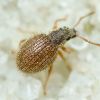
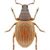
Brachysomus echinatus
Oleg Pozharov
Brachysomus (B.) echinatus (Bonsd...

31.05.2022 00:01:55 #245560
Brachysomus_3.jpg
Views: 143
Украина, Полтавская обл., Кременч...
02.05.2022
|
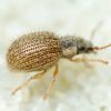
Brachysomus echinatus
Oleg Pozharov
Brachysomus (B.) echinatus (Bonsd...
31.05.2022 00:01:55 #245559
Brachysomus_2.jpg
Views: 130
Украина, Полтавская обл., Кременч...
02.05.2022
|
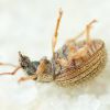
Brachysomus echinatus
Oleg Pozharov
Brachysomus (B.) echinatus (Bonsd...
31.05.2022 00:01:54 #245558
Brachysomus_1.jpg
Views: 108
Украина, Полтавская обл., Кременч...
02.05.2022
|
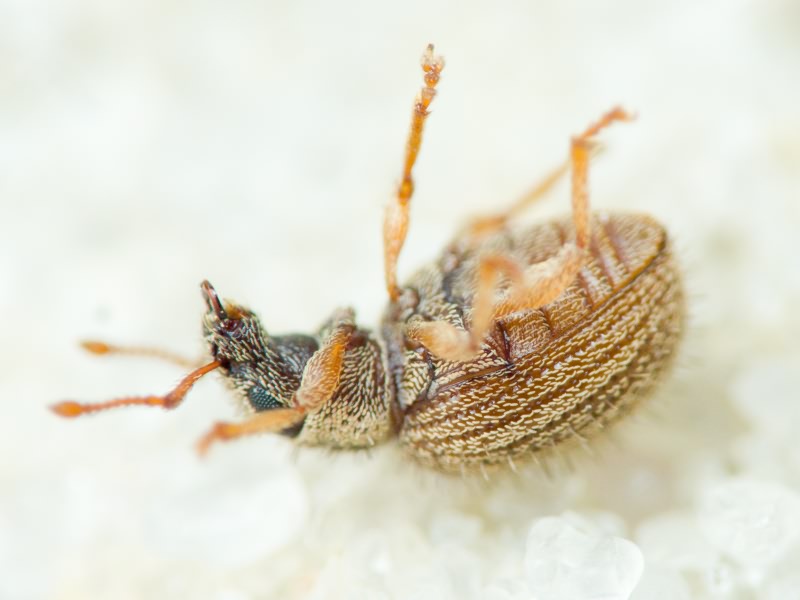
Brachysomus echinatus
Oleg Pozharov
Brachysomus (B.) echinatus (Bonsd...
31.05.2022 00:01:54 #245557
Brachysomus_4.jpg
Views: 136
Украина, Полтавская обл., Кременч...
02.05.2022
|
Brachysomus setiger
|
|
|

|
Adult
|
 |
|
Nikolai Yunakov
07.06.2008 10:30:22
06.12.2019 12:40:48 #134606
P6070269.JPG
Views: 363
Budapest, Gellért Hill
07.06.2008 (07.06.2008 10:30:22)
|
Brachysomus echinatus
barry
09.04.2018 11:32:42
10.04.2018 16:03:50 #64332
CRW_07902.jpg
Views: 474
Kharkiv, Lisopark
08.04.2018
|
Query time: 0.11 / 0.201 sec.
Query time: 0.002 / 0.204 sec.
Query time: 0 / 0.204 sec.
Online: basileus · Olexandr Kovalchuk and 394 unregistered users
BugGallery v.1.4
© UkrBIN.
PHP v. 7.2.24-0ubuntu0.18.04.17,
MySQL v. 5.7.42-0ubuntu0.18.04.1 (mysqli).


 echinatus")

 echinatus")

 echinatus")
 echinatus")
 setiger")
 echinatus")